Fluoride stains don’t require invasive dentistry to treat — enamel microabrasion, in-office bleaching, and resin infiltration can safely eliminate most discoloration while preserving your natural tooth structure. For mild cases, microabrasion uses hydrochloric acid and pumice to remove superficial stains in minutes. Moderate fluorosis responds well to combined microabrasion and 25–38% hydrogen peroxide bleaching. Severe cases may require porcelain veneers for complete masking. The right protocol depends on your fluorosis severity, and each option has specific clinical advantages worth exploring.
Key Takeaways
- Enamel microabrasion uses 6%–18% hydrochloric acid with pumice, effectively treating mild to moderate fluorosis in short, repeated application cycles.
- In-office bleaching with 25%–38% hydrogen peroxide lightens fluoride stains across multiple sessions until the desired shade is achieved.
- Combining microabrasion and bleaching targets both surface and subsurface stains, providing comprehensive treatment for moderate fluoride discoloration.
- Post-bleaching enamel sealing with low-viscosity resin composite stabilizes whitened shades and prevents stain reuptake long-term.
- Porcelain veneers effectively mask severe fluorotic stains when conservative treatments prove insufficient, offering durable, stain-resistant aesthetic results.
What Are Fluoride Stains and Why They Form on Teeth?
Fluoride stains, clinically termed dental fluorosis, develop when excessive fluoride intake disrupts ameloblast activity during enamel mineralization, producing hypomineralized enamel with subsurface porosities that scatter light and appear as white spots or brown discoloration.
Excessive fluoride disrupts enamel formation at the cellular level, leaving behind porous, light-scattering structures that appear as white spots or brown stains.
Fluoride toxicity during tooth development — typically between ages one and four — permanently alters enamel matrix protein removal, leaving porous, weakened structures beneath an intact surface layer.
You’ll notice severity ranges from faint white striations to pronounced brown staining with enamel erosion, classified using the Thylstrup-Fejerskov Index (TFI). Higher TFI scores indicate deeper structural compromise, where surface enamel breaks down post-eruption under occlusal forces.
Understanding this pathophysiology helps you make informed treatment decisions, as intervention strategies depend directly on fluorosis severity and the degree of underlying enamel integrity.
Enamel Microabrasion for Mild Fluoride Stains
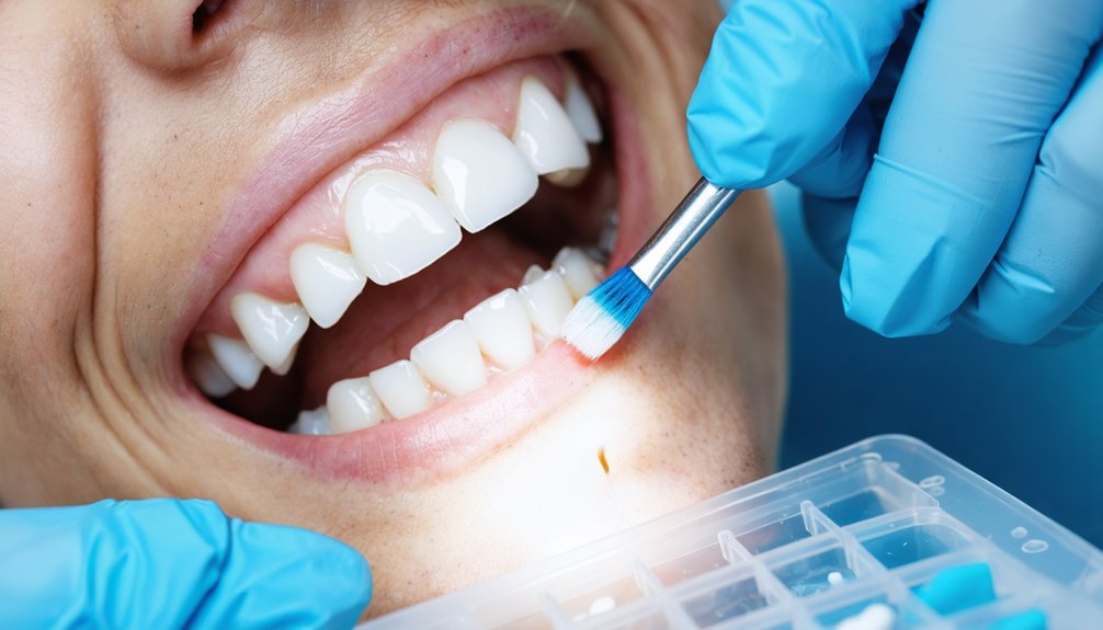
If you’re dealing with mild fluoride stains, enamel microabrasion offers a precise, clinically proven removal method using a slurry of 6% to 18% hydrochloric acid combined with silica carbide or pumice applied via a slowly rotating rubber cup in 5-second cycles, repeated up to 10 times.
You’ll find this procedure most effective for mild fluorosis cases classified at TFI scores of 1 to 3, though it’s also worth attempting for moderate cases at TFI 4.
The results are immediate and permanent, with no reported patient discomfort throughout the procedure.
Microabrasion Procedure Overview
Enamel microabrasion is the treatment of choice for mild fluorosis (TFI = 1–3) and can be attempted for moderate cases (TFI = 4).
The microabrasion technique uses 6% to 18% hydrochloric acid combined with silica carbide or pumice to remove post-eruptive calcified enamel surface material.
You’ll apply the acidic slurry using a slowly rotating rubber cup or by manually rubbing it over the affected enamel surface for 5-second intervals.
You can repeat applications up to 10 cycles per session until you achieve adequate stain reduction.
Results are immediate and permanent, with no reported patient discomfort during the procedure.
This controlled, evidence-based approach gives you predictable outcomes by mechanically and chemically eliminating superficial fluorotic discoloration without compromising deeper enamel integrity.
Ideal Fluorosis Treatment Cases
Microabrasion is ideally suited for mild fluorosis cases classified as TFI 1–3, where discoloration remains confined to the superficial enamel layer without significant structural compromise. You’re working within a treatment window where controlled enamel removal produces immediate, permanent results without restorative intervention.
For moderate cases at TFI 4, you can attempt microabrasion, though outcomes vary based on stain depth and enamel integrity.
When microabrasion alone doesn’t achieve your target shade, alternative treatment options include combining the procedure with in-office bleaching using 25–38% hydrogen peroxide, expanding your clinical flexibility.
Patient satisfaction factors strongly favor microabrasion for its single-appointment efficiency, absence of reported discomfort, and durable outcomes.
Selecting appropriate cases based on TFI classification ensures you’re maximizing procedural effectiveness while minimizing unnecessary structural tooth reduction.
Sodium Hypochlorite Bleaching for White Fluoride Stains
Sodium hypochlorite bleaching offers a clinically effective, single-appointment solution for removing white fluorosis stains. This protocol maximizes patient comfort considerations while delivering precise, controlled results as an alternative treatment option to more invasive procedures.
Follow this evidence-based protocol:
- Etch the entire tooth surface with 37% phosphoric acid for 60 seconds.
- Apply 5% sodium hypochlorite continuously for 25–30 minutes, reapplying as it evaporates.
- Rinse, dry, and re-etch for 30 seconds post-bleaching.
- Seal treated enamel with a highly penetrating clear resin composite to prevent stain recurrence.
This technique eliminates white fluorotic stains without inducing sensitivity. You maintain complete control over treatment outcomes through systematic application, ensuring predictable, long-lasting enamel whitening within a single clinical appointment.
Resin Infiltration for Fluoride Stains Bleaching Can’t Reach
When bleaching can’t fully remove fluoride stains embedded in enamel microporosities, resin infiltration offers a targeted solution by filling those voids with a light-cured dental adhesive.
Your dentist etches the enamel with 12% hydrochloric acid to expose the microcavities, then applies sodium hypochlorite to eliminate residual stain before sealing the porous surface.
A low-viscosity resin composite is then used to seal the hypomineralized enamel, preventing organic material from re-entering and ensuring long-term shade stability.
How Resin Infiltration Works
Resin infiltration works by chemically opening the microporosities within hypomineralized enamel and filling them with a light-cured dental adhesive, effectively masking subsurface fluorotic stains that bleaching agents can’t penetrate.
The protocol follows a precise sequence ensuring ideal adhesive penetration and resin durability:
- Etch enamel with 12% hydrochloric acid to remove the calcified surface layer and expose microcavities.
- Apply pure sodium hypochlorite to eliminate residual organic stains within the opened porosities.
- Infiltrate the denuded enamel with low-viscosity, light-cured resin composite for complete micropore saturation.
- Seal the hypomineralized surface to prevent organic recontamination and stabilize the corrected shade long-term.
This technique gives you precise control over stain management, targeting lesions that conventional bleaching protocols structurally can’t address.
Sealing Enamel After Bleaching
Bleaching agents penetrate enamel through surface diffusion, but fluorotic lesions with subsurface microporosities sit beyond their effective reach. Once bleaching concludes, those open pores remain vulnerable to organic stain re-uptake, compromising your results long-term.
Post-bleaching sealing closes this gap. After rinsing and drying the tooth, you etch the surface for 30 seconds, then apply a low-viscosity, light-cured resin composite. This material penetrates the hypomineralized microporosities, polymerizes within them, and locks the whitened shade permanently in place.
The clinical benefits extend beyond aesthetics. Sealing restores enamel durability by reinforcing structurally weakened fluorotic zones, reducing susceptibility to fracture and abrasion.
Regarding patient comfort, the procedure causes no sensitivity, making it a straightforward, non-invasive step that protects your investment in treatment.
Microabrasion and In-Office Bleaching for Moderate Fluoride Stains
For moderate fluoride stains that resist standalone treatments, combining microabrasion with in-office bleaching delivers a more thorough and uniform result. This protocol systematically targets both surface and subsurface discoloration through a structured sequence:
- Apply 6% hydrochloric acid microabrasion to remove calcified surface enamel containing embedded stains.
- Follow with 25–38% hydrogen peroxide or 35–40% carbamide peroxide in-office bleaching to unify tooth shade.
- Repeat bleaching sessions until your desired shade is achieved.
- Seal enamel microporosities with low-viscosity resin composite to block stain reuptake.
Alternative whitening methods alone won’t achieve comparable outcomes for TFI 4 cases. Dietary influence matters post-treatment — avoid chromogenic foods and beverages to preserve results.
This combined approach gives you precise, evidence-backed control over fluorotic discoloration management.
Dental Veneers for Severe Fluoride Stains
When microabrasion and bleaching fail to resolve extensive fluorotic discoloration, dental veneers become the indicated restorative option. These thin porcelain shells bond directly over your existing teeth, completely masking brown or severe white fluorotic stains.
Porcelain durability ensures your veneers withstand normal occlusal forces while maintaining their aesthetic integrity long-term. Their inherent stain resistance prevents future discoloration from penetrating the restoration surface.
Before placement, your clinician assesses enamel integrity. Notably, pitted enamel requires surface preparation, as rough or bumpy enamel compromises proper veneer adhesion. When substantial enamel loss exists, crowning may replace veneering as the preferred intervention.
Veneers deliver a uniform, brighter smile by fully concealing discoloration that conservative procedures couldn’t eliminate, making them a clinically sound solution for severe fluorosis cases.
Long-Term Care After Fluoride Stain Treatment
After completing fluoride stain treatment, maintaining good oral hygiene with fluoride-free toothpaste helps prevent further staining and keeps your enamel stable long-term.
Adhering to a structured maintenance protocol ensures treatment longevity and prevents reinfiltration of organic material into sealed enamel porosities.
Follow these evidence-based post-treatment directives:
- Oral hygiene: Brush twice daily using fluoride-free toothpaste to avoid disrupting resin-sealed enamel surfaces.
- Dietary habits: Limit chromogenic foods and acidic beverages that compromise sealed microporosities and accelerate stain uptake.
- Scheduled monitoring: Attend regular dental check-ups to assess resin infiltration integrity and enamel stability.
- Avoid abrasive products: Discontinue whitening toothpastes that mechanically degrade composite-sealed surfaces.
You’re protecting a clinical investment — disciplined compliance with these protocols directly determines how long your results last.
Frequently Asked Questions
Can Fluoride Stain Treatments Be Safely Performed on Young Children?
You can safely pursue fluoride stain treatments for young children, but pediatric safety demands careful treatment timing—defer microabrasion or bleaching until enamel’s fully erupted and matured, typically after age eight, to avoid iatrogenic damage.
Are Fluoride Stain Removal Procedures Covered by Dental Insurance Plans?
Insurance coverage for fluoride stain removal often falls into a “cosmetic gray area,” meaning you’ll likely bear treatment costs out-of-pocket. You should verify your plan’s specifics, as coverage varies considerably across providers and procedures.
How Long Does Each Fluoride Stain Removal Procedure Typically Take?
Each procedure varies: microabrasion takes minutes per cycle (up to 10 cycles), sodium hypochlorite bleaching lasts 25–30 minutes, and alternative whitening options differ. Natural remedies lack evidence-based timelines, so you’ll achieve faster, controlled results clinically.
Can Fluoride Stains Return After Successful Professional Treatment Is Completed?
Studies show 30% of treated cases see recurrence. Yes, stains can return if you neglect remineralization techniques and cosmetic bonding post-treatment. You’ll maintain results by sealing enamel porosities and practicing consistent oral hygiene protocols diligently.
Is It Safe to Combine Multiple Fluoride Stain Treatments Simultaneously?
You can safely combine treatments like microabrasion and bleaching, as evidence supports their chemical safety and treatment efficacy. You’ll achieve best results by sequencing procedures correctly, ensuring enamel integrity’s maintained throughout each controlled, clinically-guided step.
References
- https://dentiste-enfant.com/en/remove-stain-fluor-teeth/
- https://pmc.ncbi.nlm.nih.gov/articles/PMC11126082/
- https://www.facebook.com/groups/zerodonto/posts/10166315317545010/
- https://pmc.ncbi.nlm.nih.gov/articles/PMC6873608/
- https://journals.lww.com/jped/fulltext/2017/35030/a_comparison_of_various_minimally_invasive.14.aspx
- https://www.culligan.com/blog/how-to-remove-fluoride-from-water
- https://pmc.ncbi.nlm.nih.gov/articles/PMC4606706/
- https://www.hazendentistry.com/blog/3-ways-you-can-treat-fluoride-stains-on-your-teeth.php
- https://www.sciencedirect.com/science/article/abs/pii/S0099239906818698
- https://journals.lww.com/sjed/fulltext/2014/01010/therapeutic_management_of_dental_fluorosis__a.2.aspx