Fluoride stains respond to whitening, but only when you match the treatment to the severity of your fluorosis. Mild cases often respond well to professional whitening, while moderate-to-severe staining requires enamel microabrasion, sodium hypochlorite application, or etch-and-bleach protocols. Over-the-counter strips won’t penetrate intrinsic fluorosis lesions and can temporarily worsen visibility through dehydration. Post-treatment, you’ll need resin infiltration or fluoride varnish to protect enamel and prevent restaining. The sections ahead break down exactly which approach fits your specific case.
Key Takeaways
- Professional whitening effectively treats mild-to-moderate fluorosis by narrowing the color gap between affected and healthy enamel.
- Enamel microabrasion removes a thin outer enamel layer using acidic and abrasive agents, targeting superficial fluorosis stains effectively.
- The etch-and-bleach protocol increases enamel permeability, allowing bleaching agents to penetrate and target intrinsic fluorosis pigmentation.
- Sodium hypochlorite at 5% concentration penetrates compromised enamel within 25-30 minutes, offering a noninvasive, cost-effective whitening solution.
- Over-the-counter whitening products are ineffective for fluorosis, as the discoloration is embedded in hypomineralized enamel requiring clinical intervention.
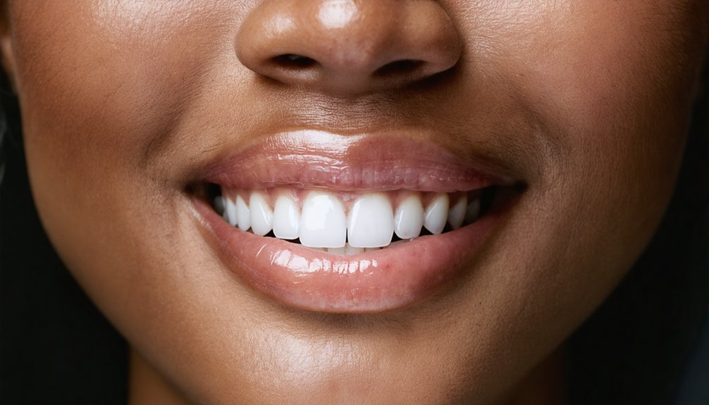
What Fluoride Stains Actually Look Like on Teeth
Fluoride stains tied to dental fluorosis don’t follow a single pattern — their appearance varies based on how much excess fluoride disrupted enamel formation during tooth development.
Mild fluoride exposure during enamel development often produces faint white mottling or chalky patches that become more visible when enamel dehydrates.
Mild fluorosis often appears as faint white patches that become more noticeable as enamel loses moisture.
Moderate cases present as broader white or light brown discoloration spread across the tooth surface.
Severe fluorosis introduces dark brown staining, surface roughness, and pitting where enamel has structurally broken down.
You’ll also notice that stain visibility can intensify temporarily after whitening procedures because dehydration amplifies contrast.
Identifying where your case falls on this spectrum matters — it directly determines which treatment options are clinically appropriate and how realistic your cosmetic outcomes will be.
Why Over-the-Counter Whitening Fails on Fluorosis
Once you understand how fluorosis affects enamel at a structural level, it becomes clearer why reaching for a store-bought whitening strip won’t solve the problem.
Fluorosis originates from excess fluoride sources during tooth development, leaving enamel hypomineralized and structurally compromised beneath its surface. Over-the-counter whitening agents work by oxidizing surface stains on intact, healthy enamel.
But fluorosis discoloration isn’t a surface deposit — it’s embedded within the enamel’s porous, defective mineral matrix.
Standard peroxide concentrations in retail products lack the depth and clinical strength to penetrate and oxidize intrinsic staining effectively.
Worse, dehydration from bleaching strips can temporarily amplify white spot visibility. Protecting enamel health requires recognizing that fluorosis demands targeted clinical intervention, not the generalized approach that consumer whitening products are designed to deliver.
Which Fluorosis Cases Respond to Professional Whitening
If your fluorosis presents as faint white mottling or small chalky spots, professional whitening offers the strongest likelihood of improvement by brightening the surrounding enamel and reducing the contrast between stained and healthy areas.
You’ll find that mild-to-moderate cases respond best because the discoloration remains relatively superficial rather than embedded in hypomineralized enamel layers.
However, if your stains appear as deep brown or gray discoloration, whitening alone won’t resolve the problem, and you’ll likely need microabrasion, bonding, or veneers to achieve meaningful cosmetic correction.
Mild Fluorosis Responds Best
When considering professional whitening for fluorosis, mild cases tend to show the most favorable results. If your fluorosis presents as faint white mottling or lightly opaque spots from early fluoride exposure, professional bleaching can meaningfully reduce contrast between affected and unaffected enamel.
As your overall tooth shade lightens, the spots become less visually prominent, aligning with realistic cosmetic expectations.
However, you’ll need to understand the mechanism: whitening doesn’t eliminate fluorosis lesions. It shifts the surrounding enamel closer in tone to the affected areas.
Dentist-supervised treatment guarantees appropriate agent selection, protecting already compromised enamel. Mild cases with smooth enamel surfaces and superficial discoloration respond predictably, whereas brown staining, pitting, or deeper hypomineralization signal that whitening alone won’t deliver sufficient correction.
Whitening Reduces Spot Contrast
Professional whitening doesn’t eliminate fluorosis lesions—it narrows the visible color gap between affected and surrounding enamel.
When excess fluoride exposure disrupts enamel development, it leaves hypomineralized zones that scatter light differently than healthy enamel. Whitening raises the overall tooth shade, reducing the contrast between those zones and adjacent surfaces.
You’ll see the best results when spots are white or lightly yellow, enamel texture is intact, and lesions cover a limited surface area.
Deeper brown stains embedded in porous enamel respond poorly because bleaching agents can’t penetrate or reverse structural hypomineralization. Some patients notice spots appear temporarily brighter post-treatment before the final shade stabilizes.
If contrast remains significant after whitening, microabrasion or bonding may be your next clinical option.
Deep Stains Need More
Mild fluorosis with small, white-to-yellow spots and intact enamel texture responds best to professional whitening—your dentist can raise the overall tooth shade enough to reduce lesion contrast without additional intervention.
However, deep brown discoloration embedded in porous or hypomineralized enamel won’t respond the same way. Standard bleaching agents can’t fully penetrate structurally compromised enamel to neutralize intrinsic pigmentation at that depth.
For those cases, your stain management strategies need to expand beyond whitening alone. Effective deep stain treatments include enamel microabrasion, sodium hypochlorite application, or etch-and-bleach protocols using hydrochloric acid followed by hydrogen peroxide.
Pitting or significant enamel breakdown may require bonding or veneers. Matching the intervention to your specific fluorosis severity is what determines whether you’ll achieve meaningful, lasting cosmetic correction.
How Enamel Microabrasion Removes Fluoride Surface Stains
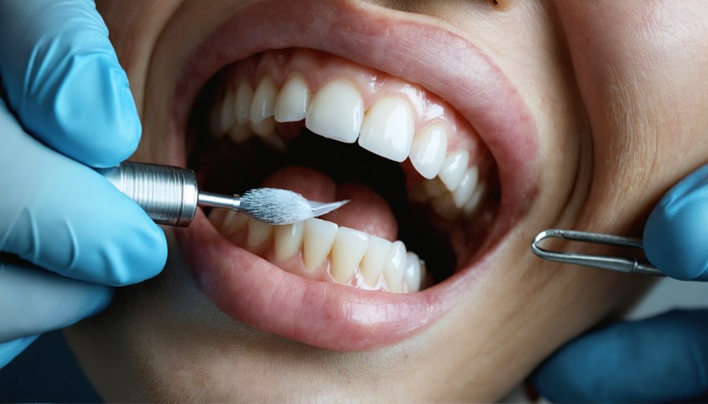
Enamel microabrasion works by removing a thin, controlled layer of the outer enamel surface using a combination of acidic and abrasive agents, typically an acid such as hydrochloric acid paired with a fine abrasive like pumice.
This mechanical-chemical process targets superficial fluorosis stains embedded in porous or discolored enamel, polishing the surface while reducing visible brown or white irregularities.
Because it preserves underlying enamel health, it’s a precise, conservative option compared to restorative alternatives.
Clinicians often combine microabrasion with professional whitening to achieve greater color uniformity post-treatment.
When evaluating your treatment options, consider that microabrasion suits shallow stains most effectively.
Deeper enamel discoloration or significant pitting may require additional interventions, so an accurate clinical assessment determines whether microabrasion alone delivers your desired outcome.
How Dentists Use Sodium Hypochlorite to Dissolve Deep Fluorosis Stains
When superficial treatments like microabrasion aren’t enough, your dentist may apply 5% sodium hypochlorite directly to stained enamel for 25 to 30 minutes, oxidizing and dissolving the organic material embedded within hypomineralized enamel lesions.
Some protocols pair this step with an etch/bleach approach—using hydrochloric acid followed by hydrogen peroxide—to target stubborn intrinsic yellow-brown discoloration more aggressively.
To prevent stain recurrence after treatment, your dentist may recommend resin infiltration, which seals the porous enamel and limits chromogen penetration going forward.
How Sodium Hypochlorite Works
Sodium hypochlorite works by oxidizing and breaking down the organic material trapped within hypomineralized enamel—the same porous, protein-rich tissue that gives deep fluorosis stains their stubborn brown or yellow color.
Its oxidation mechanisms target chromogenic proteins embedded in defective enamel, effectively neutralizing discoloration at its structural source rather than masking it superficially.
Among the core sodium hypochlorite benefits is its precision: a 5% concentration applied for 25 to 30 minutes penetrates compromised enamel without requiring mechanical removal of tooth structure.
This makes it a noninvasive, cost-effective clinical option. You’re fundamentally allowing chemistry to clear stained tissue that abrasion or conventional bleaching can’t reliably reach.
When paired with resin perfusion afterward, you further reduce the risk of chromogen re-entry into the now-cleaner enamel matrix.
The Etch And Bleach Protocol
Understanding how sodium hypochlorite oxidizes enamel-embedded chromogens sets the foundation for grasping why the etch and bleach protocol works as a complete clinical system.
In practice, dentists apply a mild acid first, using etch techniques to open hypomineralized enamel and increase surface permeability. This controlled etching allows the subsequent bleaching agent to penetrate more effectively, directly targeting intrinsic fluorosis pigmentation rather than surface deposits alone.
Bleach effectiveness improves considerably when sodium hypochlorite contacts already-opened enamel tubules, breaking down organic stain compounds through oxidation.
Clinicians typically apply 5% sodium hypochlorite for approximately 25 to 30 minutes within a single appointment. You’re achieving deeper chromogen removal without aggressive mechanical reduction, preserving enamel structure while maximizing cosmetic outcome through a low-cost, minimally invasive, evidence-supported clinical sequence.
Preventing Stain Recurrence After Treatment
Once sodium hypochlorite has oxidized and cleared embedded chromogens from hypomineralized enamel, the treated surface remains porous and vulnerable to re-staining if left unsealed.
Your post treatment care protocol should prioritize resin infiltration or fluoride varnish application immediately following the bleaching procedure, as these agents reduce chromogen penetration by occluding open enamel tubules.
For ongoing enamel protection, avoid highly pigmented foods and beverages during the initial recovery window.
Brush twice daily with a remineralizing toothpaste containing fluoride or hydroxyapatite to support enamel density.
Schedule regular professional checkups so your dentist can monitor surface integrity and intervene early if discoloration begins returning.
Combining clinical sealing strategies with disciplined home care gives you the strongest defense against stain recurrence after treatment.
When Composite Bonding Outperforms Whitening for Fluoride Stains
While whitening can reduce the contrast of mild fluorosis spots by lifting the surrounding enamel shade, it doesn’t address structural irregularities like pitting, surface roughness, or deep intrinsic discoloration embedded in hypomineralized enamel.
Whitening brightens surrounding enamel but can’t erase the pitting, roughness, or deep discoloration fluorosis leaves behind.
When those defects are present, composite bonding outperforms whitening by directly masking the compromised surface with layered resin.
Bonding techniques allow your dentist to sculpt material over affected areas, correcting both color and texture simultaneously.
Aesthetic considerations include shade matching, surface contouring, and edge blending to produce a seamless result.
Bonding works best for localized fluorosis defects rather than full-arch discoloration.
It’s minimally invasive compared to veneers, requires no enamel reduction in most cases, and delivers immediate visual correction where bleaching agents simply can’t penetrate deep enough to produce meaningful change.
Are Porcelain Veneers Worth It for Fluoride Stains?
When fluorosis causes widespread discoloration or enamel irregularities that whitening and bonding can’t adequately address, porcelain veneers offer full cosmetic coverage by capping the visible tooth surface with a thin ceramic shell.
You’ll benefit from veneers’ stain-resistant properties and their ability to mask even severe brown discoloration and surface defects in a single restorative approach.
With proper care, porcelain veneers can last 10 to 15 years, making them a durable long-term solution for fluorosis cases where conservative treatments have fallen short.
Veneer Coverage Benefits Explained
Porcelain veneers offer one of the most extensive cosmetic solutions for fluoride stains that don’t respond well to whitening or microabrasion. Among the clearest veneer advantages is full surface coverage, which masks deep brown discoloration, pitting, and enamel irregularities that bleaching can’t correct.
Your dentist bonds a thin ceramic shell directly over each affected tooth, effectively concealing structural defects beneath a stain-resistant surface. Veneer longevity is another compelling factor—well-maintained porcelain restorations typically last 10 to 15 years before replacement becomes necessary.
Unlike composite bonding, porcelain resists chromogen absorption, reducing long-term discoloration risk. You’ll need to avoid whitening after placement, as bleaching agents won’t alter ceramic and can create visible color mismatches between veneers and adjacent natural teeth.
Long-Term Veneer Durability
Investing in porcelain veneers for fluoride stains makes the most sense when you understand how long they’ll realistically hold up.
Veneer longevity depends heavily on your daily choices and clinical maintenance. Properly bonded porcelain veneers typically last 10–15 years before requiring replacement.
Key aesthetic considerations that directly impact durability:
- Avoid bleaching agents after placement — whitening products alter veneer appearance and compromise the bonding interface.
- Protect against bruxism — grinding accelerates veneer fracture, shortening their functional lifespan considerably.
- Maintain consistent dental checkups — early detection of marginal breakdown prevents costly full replacements.
You control these variables. When you manage them deliberately, veneers remain a structurally sound, stain-resistant solution for moderate-to-severe fluorosis that resists correction through bleaching or microabrasion alone.
When One Treatment Isn’t Enough for Severe Fluorosis
Severe fluorosis rarely responds to a single treatment because the discoloration is often embedded in hypomineralized enamel, making surface-level approaches insufficient on their own.
Fluorosis severity determines which treatment combinations your dentist will consider stacking together for measurable results.
A common protocol begins with enamel microabrasion to reduce surface staining, followed by a bleaching agent like sodium hypochlorite to oxidize deeper organic deposits within porous enamel.
If discoloration persists after those steps, resin infiltration or composite bonding can mask residual staining while reinforcing compromised enamel structure.
In the most resistant cases, porcelain veneers provide definitive cosmetic correction.
Each layer of treatment targets a different depth of staining, so sequencing matters.
Skipping steps or relying on whitening alone won’t address the structural irregularities driving the discoloration.
How to Protect Fluorosis-Treated Enamel From Restaining

Once fluorosis-treated enamel is restored, keeping it free from restaining requires consistent daily habits and targeted preventive care. Your enamel care routines directly determine how long your results last.
Follow these fluorosis prevention strategies to protect your investment:
- Brush twice daily with a dentist-recommended toothpaste to remove chromogens before they penetrate porous enamel surfaces.
- Limit acidic and sugary foods, which weaken enamel structure and accelerate restaining by widening microscopic entry points for discoloration.
- Schedule regular dental checkups so your dentist can monitor enamel integrity and apply resin perfusion or remineralization therapies before stains progress.
Avoiding whitening treatments after veneer placement also protects restoration appearance.
Consistent, disciplined maintenance gives you direct control over your long-term cosmetic outcomes.
Can Fluorosis Stains Return After Treatment?
Whether fluorosis stains return after treatment depends on the method used and how well you maintain your enamel afterward. Since fluorosis causes permanent changes during enamel development, the underlying structure doesn’t regenerate.
Fluorosis permanently alters enamel during development — treatment improves appearance, but the underlying structure never fully regenerates.
Treatments like microabrasion or whitening improve appearance but don’t eliminate enamel porosity, which means chromogens can re-enter over time.
Setting realistic treatment expectations matters. Whitening results fade with continued exposure to staining foods and beverages. Composite bonding can chip or discolor.
Resin infiltration reduces chromogen penetration but isn’t permanent under heavy staining conditions. Porcelain veneers offer the most durable coverage, though they require eventual replacement.
You maintain control by committing to consistent oral hygiene, limiting acidic and pigmented foods, and scheduling regular dental evaluations to monitor enamel integrity and restoration performance.
Frequently Asked Questions
Does Fluorosis Affect Children and Adults Differently in Terms of Severity?
Yes, fluorosis symptoms vary by age. You’ll notice children develop it during tooth formation, while adults retain permanent damage. Your treatment options depend on severity, ranging from whitening to veneers for lasting cosmetic correction.
Can Fluorosis Stains Be Prevented if Caught During Early Tooth Development?
Early intervention can move mountains — you can greatly reduce fluorosis severity by monitoring fluoride intake during tooth development. Making smart lifestyle choices, like limiting fluoridated water consumption, empowers you to protect your child’s enamel before damage sets in.
Is Fluorosis More Common in Certain Geographic Regions With Higher Fluoride Levels?
Yes, you’ll find fluorosis more prevalent in regions with naturally high fluoride sources in groundwater. Geographic variation drives exposure risk, and health policies alongside public awareness campaigns help you understand and manage your local fluoride levels effectively.
Does Dental Insurance Typically Cover Cosmetic Treatments for Fluorosis Stains?
Your dental insurance typically won’t cover cosmetic treatments for fluorosis stains, as insurers classify them as elective. You’ll need to verify your plan’s policy directly, since coverage varies considerably between providers.
Can Fluorosis Stains Affect the Back Teeth as Well as Front Teeth?
Yes, fluorosis impact doesn’t spare your back teeth. You’ll find discoloration, pitting, and enamel irregularities affecting molars and premolars equally, since excess fluoride exposure during development systematically targets all forming teeth, not just front teeth.
References
- https://www.hazendentistry.com/blog/3-ways-you-can-treat-fluoride-stains-on-your-teeth.php
- https://pmc.ncbi.nlm.nih.gov/articles/PMC4606706/
- https://www.ritchiedentalgroup.com/blog/expert-dentist-in-marble-falls/
- https://www.dentistsatorenco.com/blog/white-spots-dental-fluorosis-treatment/
- https://www.gilaridgedental.com/why-do-some-people-get-white-spots-after-whitening/
- https://www.brookwooddentistry.net/how-to-get-rid-of-white-stains-on-teeth/
- https://www.medicalnewstoday.com/articles/322112
- https://www.colgate.com/en-us/oral-health/teeth-whitening/teeth-stain-removal-types
- https://www.ruidosodentist.com/how-to-get-rid-of-white-stains-on-teeth/
- https://crispinchangdds.com/blog/5-everyday-oral-hygiene-tips-to-whiten-teeth/